Plant embryology is the branch of biology that deals with the formation, structure, growth, and development of embryos produced through the union of male and female gametes.
Fundamental terminology of plant embryology:
- Embryo: The embryo is a miniature plant within a seed produced due to the union of male and female gametes, resulting in the development of a zygote.
- Microsporophyll: Every member of androecium is called the stamen, which is known as microphyll.
- Megasporophyll: Every member of gynoecium is called carpel, which is known as megasporophyll.
- Spore (n): The reproductive cell of higher plants is the unit of cytoplasm and can fuse with the gamete. There are two types of spore: Male spore (microspore/pollen) and Female spore or megaspore.
- Male spore (microspore/pollen): A haploid spore originated from the meiotic division of the pollen mother cell or microspore mother cell.
- Female spore or megaspore: The giant spore rises from a female gametophyte in which an egg is produced.
- Gamete (n): It is the unit of cytoplasm that can fuse with other gametes. There are two types of gamete –Male gamete or microgamete (n), Female gamete or mega gamete (n).
- Sporophyte: Spore bearing plant is called Sporophyte.
- Gametophyte: Gamete bearing plant is called gametophyte.
- Pollen mother cell (PMC) or microspore mother cell (2n): A diploid (2n) cell gives through meiosis 4 haploid microspore.
- Megaspore mother cell (MMC): A diploid (2n) cell undergoes two divisions to produce 4 haploid megaspores.
- Microsporangium: The pollen sac in which pollen or male spore are developed and stored is called microsporangium.
- Megasporangium: The ovule in which female spores or megaspores are developed and stored is called megasporangium.
- Sporogenesis: It is the formation process of young spores within the sporangium. They are two types.
- Pollination: Pollination is the transference of pollen grains from the anther of a flower to the stigma of the same flower or of another flower of the same or sometimes allied species.
- Fertilization: Fertilization is the process by which male and female gametes are fused together, initiating the development of a new organism.
- Double fertilization: Double fertilization is the union of one male gamete with an egg and the other male gamete with the secondary nucleus of the same embryo sac.
- Triple Fusion: Triple fusion is the fertilization of two polar nuclei in the embryo sac with a single sperm cell and produces the triploid endosperm.
- Parthenogenesis: A form of asexual reproduction where the offspring develops from the egg or female gamete without the prior fertilization from the male gamete.
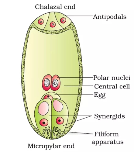
The structure of mature embryo sac:
The embryo sac lies embedded in the nucellus towards the micropylar end and consists of the following parts:
- Synergids: Usually, the pair of synergid cells are apparently equivalent and similar. The upper ends of this pair are attached to the embryo sac wall while the lower ends of the cells produce into the sac cavity. Here, the lower round end of each synergid has a large vacuole. Here the walls of synergids are made of cellulose.
- The oosphere: The egg apparatus comprises two parts, two synergids and an egg (oosphere); the former has been just described above. Typically this oosphere is found attached to the synergids. At the time of fertilization, it seems somewhat large in size. This oosphere is supposed to be metabolically inactive, and so it receives its nourishment from the synergids.
- The central cell: It is the largest cell of the embryo sac. The nuclei of it are termed polar nuclei. The number of polar nuclei in an embryo sac is not definite but varies from species to species. The polar nuclei unite before, during, or after the pollen tube entry into the embryo sac, and the product is called the secondary nucleus.
- The antipodal cells: Short-lived cells have a significant variation in size, number, and durability. Gradually these cells are found in a group of three cells separated by delicate cell walls.
Microsporogenesis:
Microsporogenesis is the formation process of the pollen grain, i.e., microspore and its development within the microsporangium.
- Making a cross-section of a very young developing anther shows a mass of homogeneous meristematic tissue surrounded by the epidermis.
- With further development, the anther becomes 4 lobed at each lob, and hypodermal cell/cells become conspicuous and differentiated. The cell is larger-sized with a prominent nucleus. This cell is called the archesporium (2n).
- These archesporial cells divide to form a primary parietal layer toward the outside and a primary sporogenous layer toward the inner side.
- The primary parietal layer mitotically divides and produces 3 – 5 layers of anther wall.
- The primary sporogenous layer undergoes mitotic division to form sporogenous tissue.
- This sporogenous tissue gives rise to the Microspore mother cell (2n) or may undergo further division and produce the Microspore mother cell (2n).
- Finally, the Microspore mother cell (2n) undergoes divisions and usually produces haploid microspore, or pollen grain arranged tetrahedrally.

Gametogenesis:
It is the process of gamete formation from spore and the duration, called gametogenesis.
Microgametogenesis:
It is the formation process of male gamete from the microspore produced by microsporogenesis.
Microspore or pollen grain is the first cell of the male gametophyte which contains only one haploid nucleus. Each pollen grain is covered with two epidermises named intine and exine. The outer epidermis is thick, hard, and commenting cuticle called exine. In various places of exile, there is a very thin pore; these are called germ pores.
- At first, the nucleus of the microspore undergoes unequal divisions and forms a generative nucleus and large vegetative nucleus.
- Microspore becomes fatty absorbing sticky sap of stigma, and pressure is created inside the microspore. At this time, in the place of germ pore, exine breaches, and intine increases in tubular ways. It is called a pollen tube. If a pollen tube is created, the nucleus entering it stays on the top part. Then the nucleus enters inside.
- Initially, the generative nucleus remains to lie with intine. The pollen tube gradually rises through stigma, and later on, the generative nucleus divides mitotically and produces two male gametes. After producing the male mate gamete, the vegetative nucleus degenerates.

Megasporogenesis:
Megasporogenesis is the formation process of MMC (Megaspore Mother Cell) within the megasporangium (ovule) from the megaspore.
The longitudinal section of a very young developing ovule shows the archesporial tissue at the hypodermal origin. In general, one cell of the nucellus situated directly below the epidermis becomes more conspicuous, having a larger size, dense cytoplasm, and prominent nucleus called the primary archesporium cell. It may divide to form a primary parietal cell and a primary sporogenous cell or function directly as the megaspore mother cell.
The primary sporogenous cell may function as the megaspore mother cell (2n) without further divisions. Megaspore mother cell (2n) undergoes meiotic divisions to form a linear tetrad of four cells. Usually, the outer three degenerate, and the inner one becomes a functional megaspore, giving rise to the embryo sac later on.

Megagametogenesis:
It is the process of formation of female gamete from the functional megaspore. The embryo sac formation from the mother cell shows a lot of variation. When 4 megaspores are formed from the megaspore mother cell (MMC), and only one of the four gives rise to the embryo-sac, the process is called the monosporic process, and it occurs 75% in the plant.
Megaspore is the first female gametophyte cell known as embryo sac. The functional megaspore increase in size and develops into a simple female gametophyte or embryo sac.
The nucleus of the functional megaspore is divided by mitosis forming 8 nuclei. At first, the nuclei within the embryo sac are arranged in two groups of 4 nuclei at the micropylar and chalazal end.
After that, one nucleus from each group then migrates to the middle of the embryo sac and fuses to form a diploid (2n) nucleus, called the secondary or polar fusion nucleus.
The 3 nuclei remaining at the micropylar end become surrounded by cytoplasm and form the egg apparatus. The middle layer cell of the egg apparatus is known as the egg or ovum, flanked by two synergids. The other 3 cells take a position opposite the egg cell, known as antipodal cells. The egg is known as a megagamete or female gamete.

Development of endosperm (Dicot plant):
The endosperm is an important tissue that is the embryo’s primary and future food source. A male gamete fertilizes the polar nuclear in gametic fusion to form the primary endosperm nucleus (3n). After fertilization, it goes into some resting period. Afterward, this endosperm nucleus will form endosperm by breaking the resting period with mitotic division. There are four ways for the development of endosperm.
Nuclear type: In this type, the endosperm nucleus divides repeatedly, but the cell wall or cytoplasm will not divide. As a result, several free nuclei appear in the embryo sac. As division progress, the nuclei become pushed more or less towards the periphery, so the center is occupied by a large vacuole.
Each nucleus is surrounded by cytoplasm and ultimately produces individual cells (Rarely two or three or more produce individual cells). Then the cell wall formation starts from embryosac and proceeds towards the center to fill the center at last nodule / matured endosperm produced.
Cellular type: In this type, as the endosperm nucleus divides, there is a corresponding formation of the cell wall around each nucleus.
The cell wall divides as many times as the endosperm nucellus divides; as a result, the embryo sac becomes divided into several chambers.
At first mitotic division, the wall divides transversely. But afterward, the division may be transversely or oblique or longitudinally, and at last, this will be produced a matured endosperm.
Global type: It is intermediate between the nuclear type and cellular type. The first division of the endosperm and nucleus results in embryo sac formation into two chambers: the micropylar side.
Several free nuclear divisions take place in the micropylar chamber, but in the chalazal chamber, either the nucleus remains undivided or undergoes a few divisions and then degenerates. Finally, the nuclei of the micropylar side produce the entire endosperm.
Ruminate type: Ruminate means chewing. The ruminant condition result due to irregular growth of seed code and endosperm.
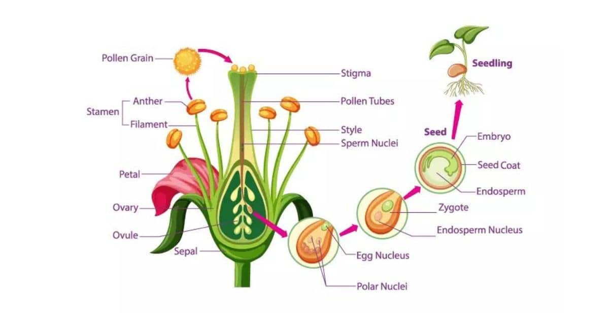
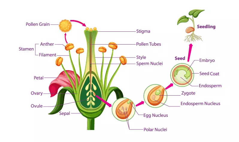
Process of fertilization in plant:
Fertilization is the process by which male and female gametes are fused together, initiating the development of a new organism.
When the pollen tube reaches the ovule, it enters the ovule generally through micropyle or penetrates through the integuments and fuses with the embryo sac. The wall between the two dissolves at the fusion point and two male gametes are ejected into the embryo sac. One male gamete fuse with the egg and thus fertilizes the egg.
The fertilized egg secrets walled around it and converted to a diploid (2n) zygote. The other male gamete in the embryo sac fuses with the secondary nucleus forming a triploid endosperm nucleus. The fusion of the male nucleus with the secondary nucleus is termed double fertilization. The zygote develops into the embryo, and endosperm tissue is formed from the endosperm nucleus. It is used as food for the developing embryo.
Double fertilization:
Fertilization is the process by which male and female gametes are fused together, initiating the development of new organism fertilization occurs twice.
One of the two male gametes of the pollen tube fuses with the ovum of the embryosac. The other gamete fuses with the definitive nucleus. This process is called fertilization.
Double fertilization is the union of one male gamete with egg/ovum and the other male gamete with the secondary nucleus of the same embryo sac. It was first discovered by NawaSchin in 1898 by Lilium and Fritillaria.
This amazing discovery attracted great attention. Many investigators soon established that double fertilization is a universal occurrence among angiosperm. The significance of double fertilization is not clearly understood.
The fusion of male gametes with the ovum results in the formation of the embryo, and the definite nucleus results in the formation of the endosperm.
Entry of pollen tube into embryo sac:
After entering the wall of the embryo sac, the pollen tube during its entry into the embryo sac passes the following ways.
- Porogamy: It is the normal method. This process occurs in the maximum plant. When the pollen tube enters the embryo sac through the micropyle. e.g., Mango
- Chalazogamy: When the pollen tube entry into the embryo sac through the base of the ovule. e.g., Casuarina.
- Monogamy: When the pollen tube enters the embryo sac through the integuments. e.g., Sweet gourd
Types of the embryo sac in angiosperm:
Based on the number of megaspore nuclei, the female gametophyte of angiosperm may be grouped into 3 types:
- Monosporic: Only one takes part in female gametophyte development out of four megaspores in the monosporic type.
- Biosporic: Two megaspore nuclei take part in the development of female gametophytes.
- Tetrasporic: All four megaspore nuclei participate in the development of female gametophytes.
Classification of embryo sac based on early development:
Johansen recognized 6 types of embryos developed among the angiosperm, the basis of basal and terminal cells.
- Piperade type
- Onagrad or crucifer type
- Instead type
- Caryophyllad type
- Solana type
- Chenopoded type
1. Piperad type – eg: Peperomia, Balanophora
A. The zygote is divided by a more or less vertical wall.
B. The zygote is divided by a transverse wall.
I. The terminal cell is divided by a vertical wall forming an inverted ‘T’ shaped proembryo.
- The basal cell plays little or no part in the embryo’s development.
2. Onagrad type – e.g., Onagraceae, Liliaceae family.
- Both basal and terminal cells contribute to the formation of the embryo.
3. Asterad type – eg: compositae family.
II. The terminal cell divides by a transverse wall forming a straight embryo.
- Basal cell plays little part in the further development of the embryo. The basal cell undergoes no further division but becomes a large suspensor cell.
4. Caryophyllad type – e.g., Caryophyllaceae.
- Basal cell forms suspensor of two or more cell
5. Soland type – eg: Solanaceae, Papavaraceae family
- Both basal and terminal cells contribute to the formation of the embryo.
6. Chenopoaded type – e.g., Chenopodiaceae family.
Megasporangium:
The ovule in which female or megaspores are developed and stored is called megasporangium.
Structure of anatropous ovule / Megasporangium:
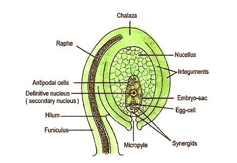
Anatropous is most common among angiosperms. It consists of the following part.
- Funicle: The ovule is a rounded structure attached to the placenta by a stalk known as a funicle.
- Hilum: The place of attachment of the funicle to the body of the ovule is known as the hilum.
- Chalaza: The basal region of the ovule, where integuments arise, is known as chalaza.
- Raphe: In anatropous ovules, the funicle extends above along the body of the ovule to form a ridge known as raphe.
- Nucellus: The main body of the ovule is called the nucellus.
- Integuments: Nucellus is surrounded by two coats termed called integuments. It may be one (unitegmic) or two (bitegmic).
- Micropyle: The extends well beyond the nucellus to form a narrow opening called the micropyle.
- Embryo sac: A large, oval cell lying in the nucellus towards the micropylar end called the embryo sac. It consists of egg cell and synergid. These are known as egg apparatus and they have two parts namely polar nucleus/secondary nucleus and antipodal cell.
Ovule:
Ovule is defined as an integument megasporangium. It encloses the embryo sac, which is the female gametophyte of angiosperm.
Types of ovule :
- Atropous or orthotropous: The ovule is straight so that the micropyle lies on the same vertical axis with the funicle and chalaza. e.g., Polygonum, Piper.
- Anatropous: In this type, the body of the ovule becomes completely inverted so that the micropyle and hilum come to lie very close to each other. The micropyle and chalaza lie on the same vertical axis but not the funicle. e.g., Castor, Helianthus.
- Campylotropous: When the ovule is curved so that the micropyle and chalaza do not lie on the same straight line. It is called campylotropous. e.g., Pea, Mustard.
- Amphitropous: When the ovule’s curvature is so pronounced that the embryo sac bends like a horseshoe, the ovule is called amphitropous. e.g., Poppy, etc.
- Hemianatropous: In this type, the nucellus and integuments lie more or less at right angles to the funicle. e.g., Ranunculus
- Circinotropous: In this type, the nucellar protuberance is at first in the same line as the axis, but the rapid growth on one side makes it anatropous. The curvature continues until the ovules turn entirely, with the micropylar end again pointing upward. e.g., Opuntia

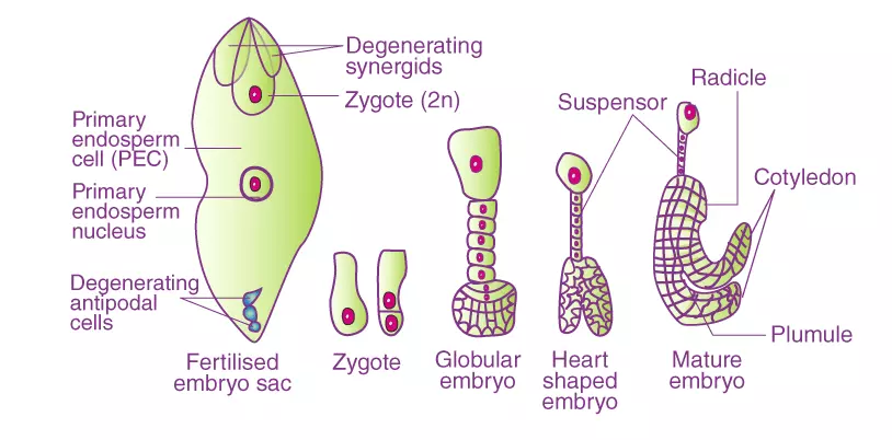
Development of embryo:
After fertilization, the fertilized egg is called a zygote or oospore, which develops into an embryo. The fertilized egg or zygote usually represents the formation of the embryo. The zygote is a unicellular system that gives a multicellular embryo with developed or different organs. Finally, the embryo forms an adult plant.
Before it actually enters into the process, the oospore undergoes a rest, which may vary from a few hours to a few months. Generally, the zygote divides immediately after the first division of the primary endosperm nucleus, but sometimes it divides earlier than the primary endosperm nucleus. In angiosperm, the zygote is located in the micropylar region of the embryo.
With the development of endosperm, the zygote develops the embryo. In all angiosperms, the zygote divides and forms two cell proembryo (the embryo is called proembryo after 1st division). In most plants, the first wall between the two cells is transverse, while in a few cases, the first wall is more or less vertical (the first division of the zygote is always followed by a wall – formation resulting in a two-celled proembryo.
Polyembryony:
It is defined as the production of two or more embryos within one ovule. It is defined as the occurrence of more than one embryo in a single seed. Besides the seed egg, the other constituents of cells or embryosac with or without fertilization produced embryos.
Outside the embryo sac, the activity of one or more cells also develops accessary embryos. In addition to the zygotic embryo, sexual or asexual non-zygotic embryos may also develop.
Apomixis:
A means no; mixis means mixture, which means without mixture. According to Winkler, apomixis may be defined as substituting sexual reproduction for an asexual process that does not involve any nuclear fusion.
Apomixis is a modified form of reproduction in which seeds are formed without fertilization or gamete fusion. Apomixis is a form of asexual reproduction because there is no male and female gametes union. The fruits developed by apomixis are called parthenocarpic fruits.